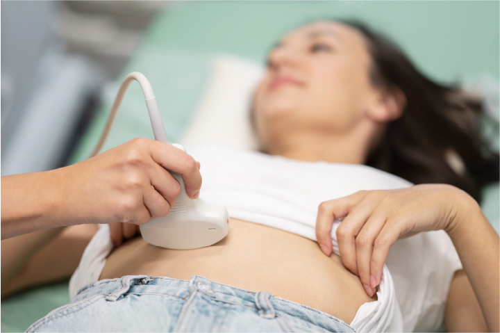
Ultrasonografi (USG)
Ultrasonografi abdomen dan pelvis merupakan uji pencitraan non-invasif yang menggunakan gelombang suara frekuensi tinggi untuk membuat gambar terperinci dari struktur di dalam area tubuh tersebut. Umumnya digunakan untuk mengevaluasi organ dan jaringan di daerah abdomen dan pelvis untuk berbagai kondisi medis.
Tujuan
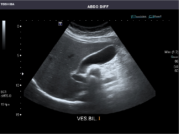
USG-ABDOMEN
USG digunakan untuk melihat dan mengidentifikasi kondisi organ seperti hati, kantong empedu, limpa, pankreas, dan ginjal. Orang tersebut harus berpuasa selama 6 jam sebelum menjalani prosedur. Konsumsi air putih dan obat-obatan diperbolehkan.
USG-PELVIS
USG pelvis memberikan informasi kepada dokter mengenai kondisi sistem reproduksi dan saluran kemih seseorang. Untuk mempersiapkan prosedur, orang tersebut harus minum banyak air setidaknya satu jam sebelum USG agar organ pelvis (kandung kemih, prostat, rahim, dan ovarium) dapat terlihat lebih jelas.
Cara Melakukan Tes
Persiapan
Posisi
Pemindaian

REMS (Radiofrequency Echographic Multi Spectrometry)
Teknologi bebas radiasi untuk menilai kepadatan tulang dan kualitasnya pada vertebra lumbar dan femur proksimal melalui pendekatan revolusioner untuk mengkarakterisasi mikroarsitektur tulang menggunakan sinyal frekuensi radio yang diperoleh selama pemindaian ultrasonografi sederhana.
Metode ini mengatasi semua keterbatasan utama DXA (Dual-Energy X-ray Absorptiometry) dan QUS Ultrasound). Teknik diagnostik non-invasif yang digunakan untuk menilai kesehatan tulang, khususnya untuk mendiagnosis osteoporosis dan mengevaluasi risiko patah tulang.
Tujuan
PEMERIKSAAN UNTUK OSTEOPOROSIS
Terutama pada wanita pascamenopause dan orang dewasa yang lebih tua.
PEMANTAUAN KESEHATAN TULANG
Pada pasien dengan osteoporosis atau osteopenia yang diketahui.
PENILAIAN RISIKO PATAH TULANG
Untuk individu dengan faktor risiko patah tulang.
EVALUASI KESEHATAN TULANG PADA POPULASI KHUSUS
Seperti mereka yang menjalani terapi steroid jangka panjang atau dengan penyakit kronis yang memengaruhi metabolisme tulang.
Cara Melakukan Tes
- Pasien biasanya berbaring dengan nyaman di meja pemeriksaan
- Area yang akan diperiksa, biasanya tulang belakang lumbar atau pinggul, biasanya akan diekspos.
- Gel berbahan dasar air dioleskan ke kulit di atas area yang akan dipindai untuk memfasilitasi penghantaran gelombang ultrasonik
- Pemindaian ultrasonik hanya membutuhkan waktu 80 detik untuk tulang belakang dan 40 detik untuk tulang paha.
- Perangkat lunak REMS menganalisis data ultrasonik untuk menilai kepadatan mineral tulang (BMD) dan sifat mikroarsitektur tulang.
- Mengevaluasi berbagai parameter yang penting untuk menentukan kekuatan tulang dan risiko patah tulang.
- Laporan terperinci dibuat, yang menunjukkan kepadatan tulang dan memberikan penilaian osteoporosis atau osteopenia jika ada.
Pemindaian Hati Berlemak
Pemindaian Hati Berlemak adalah prosedur non-invasif yang digunakan untuk menilai kesehatan hati, khususnya dalam mendeteksi dan memantau hati fibrosis dan steatosis (perlemakan hati).
Dengan menggunakan teknologi transient elastography untuk mengukur kekakuan dan kandungan lemak jaringan hati. Mesin ini dinilai cepat, tidak menimbulkan rasa sakit, dan dapat diulang, sehingga memungkinkan pemantauan kesehatan hati secara teratur pada pasien dengan kondisi hati kronis.
Tujuan
1. Diagnosis Penyakit Hati Berlemak: Penyakit Hati Berlemak Non-Alkohol (NAFLD) atau Penyakit Hati Beralkohol (ALD)
2. Menilai Keparahan Steatosis: Memberikan ukuran kuantitatif kandungan lemak hati
3.
Deteksi Fibrosis Hati: menilai kekakuan hati, yang berkorelasi dengan fibrosis. Ini penting dalam mengidentifikasi perkembangan dari steatosis sederhana ke kondisi hati yang lebih parah seperti steatohepatitis non-alkohol (NASH), fibrosis, atau sirosis.
4. Deteksi dan Pencegahan Dini: Deteksi dini lemak hati dan fibrosis memungkinkan dilakukannya intervensi tepat waktu yang dapat mencegahnya perkembangan penyakit hati yang lebih parah seperti sirosis atau kanker hati.
Cara Melakukan Tes
1. Persiapan:
- Pasien biasanya berbaring telentang dengan lengan kanan diangkat di atas kepala untuk memperlihatkan sisi kanan perut tempat hati berada.
- Biasanya tidak diperlukan persiapan khusus, meskipun mungkin disarankan untuk berpuasa beberapa jam sebelum pemindaian.
2. Perangkat dan Metode::
- Perangkat FibroTouch menyerupai mesin USG dengan probe khusus.
- Probe memancarkan getaran frekuensi rendah dan gelombang ultrasonik tanpa rasa sakit ke dalam hati.
- Gelombang ini menciptakan gelombang geser pada jaringan hati, dan kecepatan perjalanan gelombang ini berkorelasi dengan kekakuan jaringan.
3. Pengukuran:
- Pengukuran Kekakuan Hati (LSM): Output utama adalah nilai kekakuan hati, yang dinyatakan dalam kilopascal (kPa). Nilai kekakuan yang lebih tinggi menunjukkan