Elektrokardiogram (EKG)
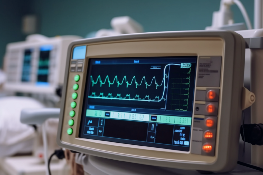
EKG adalah tes medis yang mengukur aktivitas listrik jantung. Ini adalah tes yang sederhana, aman, dan tidak menyakitkan. Tidak ada risiko tersengat listrik.
Tujuan
- Mendiagnosis kondisi jantung: aritmia, serangan jantung, dan masalah jantung lainnya.
- Memantau kesehatan jantung: Membantu memantau kesehatan jantung dari waktu ke waktu, terutama pada pasien dengan kondisi jantung yang sudah ada.
- Penilaian praoperasi: Digunakan sebagai bagian dari evaluasi praoperasi untuk memastikan jantung dapat menangani tekanan operasi.
Cara Melakukan Tes
Persiapan
Pasien berbaring, dan elektroda dipasang di dada, lengan, dan kaki.
Perekaman
Mesin EKG merekam aktivitas listrik jantung.
Analisis
Jejak EKG yang direkam dianalisis oleh profesional perawatan kesehatan.

ECHO
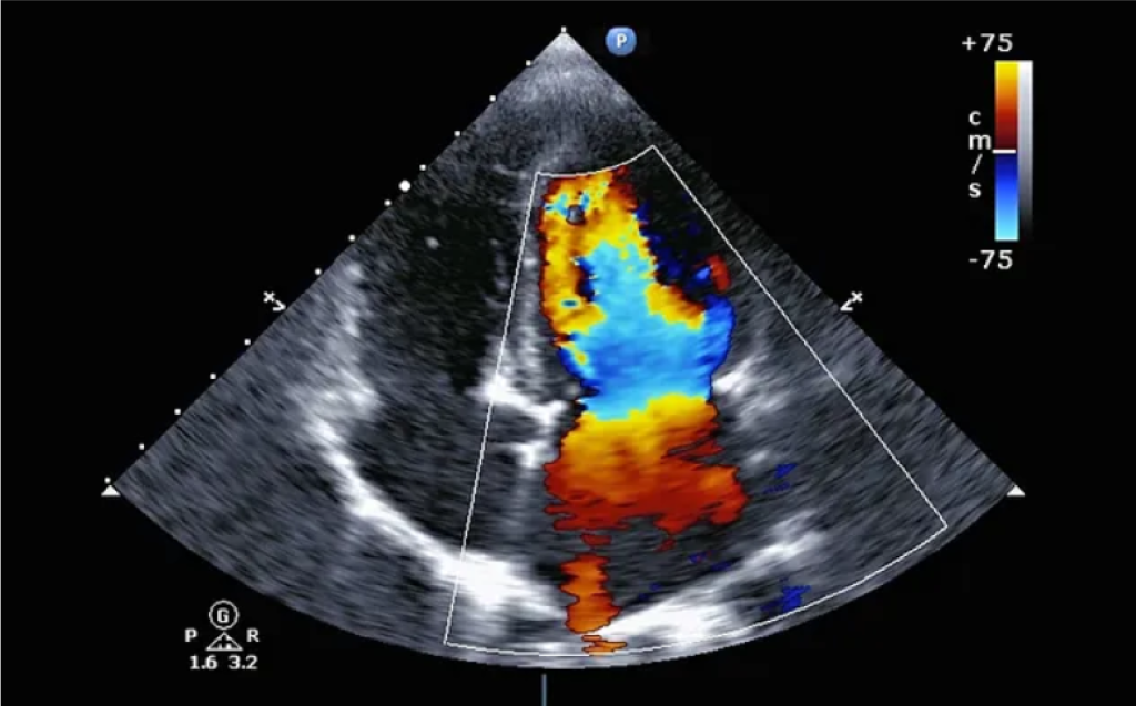
Ekokardiogram, yang sering disebut sebagai “echo,” adalah tes diagnostik yang menggunakan gelombang ultrasonik untuk membuat gambar jantung. Prosedur non-invasif ini memberikan informasi terperinci tentang struktur dan fungsi jantung.
Tujuan
- Menilai fungsi jantung: Mengevaluasi ukuran, bentuk, dan pergerakan bilik dan katup jantung.
- Mendeteksi kondisi jantung: Mengidentifikasi kelainan seperti penyakit katup, cacat jantung, kardiomiopati, dan efusi perikardial
- Memantau kondisi jantung yang sedang berlangsung: Melacak perkembangan penyakit jantung dan efektivitas pengobatan.
- Evaluasi pra-bedah: Menilai fungsi jantung sebelum operasi.
Jenis-jenis Ekokardiografi
- Ekokardiogram Transtoraks (TTE): Ini adalah jenis ekokardiogram yang paling umum dan dilakukan dengan menempatkan transduser pada dinding dada untuk menangkap gambar jantung dari luar tubuh.
- kokardiogram Transesofageal (TEE): Dalam beberapa kasus, TEE dapat dilakukan, yang melibatkan penyisipan probe khusus ke dalam esofagus untuk mendapatkan pandangan jantung yang lebih dekat dan jelas.
Ini sering digunakan untuk penilaian yang lebih rinci. - Ekokardiogram Stres: Tes ini menggabungkan ekokardiografi dengan latihan atau pengobatan untuk mengevaluasi bagaimana jantung berfungsi di bawah tekanan.
Cara Melakukan Tes
- Tes gema dilakukan oleh profesional perawatan kesehatan yang terlatih
- Tes ini tidak menimbulkan rasa sakit dan tidak memiliki efek samping.
- Biasanya memakan waktu kurang dari satu jam.
- Selama tes
- Pasien berbaring di tempat tidur di sisi kiri atau belakang.
- Sonografer akan meletakkan gel khusus pada probe dan menggerakkannya di atas dada pasien.
- Gelombang suara akan berjalan melalui probe dan mengambil gambar jantung dan katup.
- Tidak ada sinar-X yang akan digunakan dan tidak akan terpapar radiasi.
- Pergerakan jantung dapat dilihat di layar video.
- Terkadang probe harus lebih dekat ke jantung pasien untuk memberikan gambaran yang lebih jelas. Jika hal itu terjadi, mungkin diperlukan tes khusus yang disebut kardiogram transesofageal (TEE), tes ini untuk mencari tanda-tanda infeksi (endokarditis), bekuan darah (trombus), atau struktur atau koneksi abnormal lainnya.
- Selama tes ini:
- Puasa diperlukan sebelum tes
- Pasien mungkin mendapatkan obat untuk membantu rileks dan cairan mati rasa disemprotkan di bagian belakang tenggorokan.
- Kemudian, seorang ahli jantung akan dengan lembut memasukkan tabung dengan probe di ujungnya ke tenggorokan dan ke dalam kerongkongan.
- Gelombang suara akan menciptakan gambar seperti yang dijelaskan di atas.
- Setelah tes selesai, ahli jantung akan dengan lembut menarik probe keluar.
- Mungkin merasa tidak nyaman dan perlu batuk.

Tes Stres
Tes stres (juga dikenal sebagai tes latihan jantung) digunakan untuk mengukur respons jantung terhadap aktivitas fisik dalam lingkungan yang terkendali. Tes stres menilai seberapa baik jantung menangani pekerjaan dengan memantau denyut jantung, tekanan darah, elektrokardiogram, dan kadar oksigen saat pasien beristirahat dan selama aktivitas fisik.
Tujuan
- Mendiagnosis Penyakit Arteri Koroner (CAD): Mengidentifikasi penyumbatan pada arteri koroner.
- Mengevaluasi Irama Jantung: Mendeteksi aritmia yang mungkin terjadi selama latihan.
- Menilai Fungsi Jantung: Mengevaluasi seberapa baik jantung memompa darah di bawah tekanan.
- Memandu Rencana Perawatan: Membantu menentukan efektivitas perawatan saat ini atau kebutuhan untuk intervensi baru.
- Mengembangkan Program Latihan: Memberikan informasi untuk pedoman latihan yang aman untuk rehabilitasi jantung.
Cara Melakukan Tes
1. Persiapan:
- Kenakan pakaian dan sepatu yang nyaman dan sesuai untuk latihan.
- Hindari makanan berat, kafein, atau merokok selama beberapa jam (2 hingga 3 jam) sebelum tes.
- Beri tahu dokter tentang semua obat yang sedang dikonsumsi
2. Selama Tes:
- Elektroda akan dipasang di dada untuk memantau aktivitas listrik jantung (EKG).
- Manset tekanan darah akan dipasang di lengan.
- Mulailah berolahraga di atas treadmill, tingkatkan intensitas secara bertahap, pasien akan diminta berjalan di atas treadmill dengan kecepatan cepat selama sekitar 15 menit dan berusaha mencapai detak jantung tertentu (berdasarkan usia). Setiap 3 menit, treadmill akan menambah kecepatan dan kemiringan.
- Staf medis terlatih akan mengawasi pasien setiap saat dan memastikannya dilakukan dengan aman.
- Denyut jantung, tekanan darah, dan EKG akan dipantau selama tes.
3. Setelah Tes:
- Pasien akan dipantau untuk waktu yang singkat saat detak jantung dan pernapasan kembali normal.
